
Label the histology of the ovary using the hints provided. – Embarking on an exploration of the intricate histological landscape of the ovary, this discourse delves into the defining characteristics, structural organization, and functional significance of its cellular components, providing a comprehensive understanding of this vital reproductive organ.
Unveiling the dynamic processes of follicular development, corpus luteum formation, and the spectrum of ovarian cysts and neoplasms, this analysis illuminates the histological underpinnings of ovarian physiology and pathology.
Ovarian Histology Overview

Ovarian histology refers to the microscopic study of the ovary, a female reproductive organ responsible for producing eggs and hormones. The ovary is a complex organ with a unique structure and organization, composed of various cell types and layers. Understanding ovarian histology is crucial for comprehending its function, development, and potential pathologies.
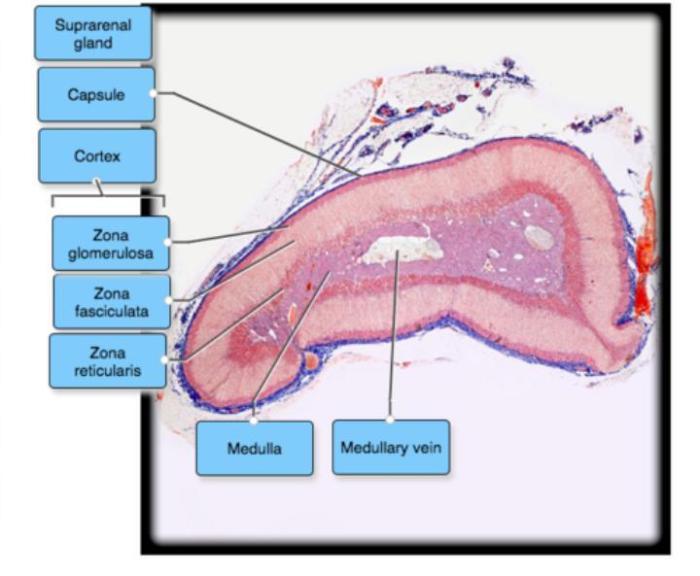
The ovary consists of an outer layer of surface epithelium, a cortex containing ovarian follicles, and a medulla with blood vessels and connective tissue. The surface epithelium is a single layer of cuboidal cells that covers the ovary and functions as a protective barrier.
The cortex is the functional zone of the ovary, housing ovarian follicles at different stages of development. The medulla provides structural support and contains blood vessels, nerves, and lymphatic vessels.
| Component | Histological Features | Staining Characteristics | Function |
|---|---|---|---|
| Surface epithelium | Single layer of cuboidal cells | Eosinophilic cytoplasm | Protection |
| Cortex | Contains ovarian follicles | Follicles stain with hematoxylin and eosin | Production of eggs |
| Medulla | Contains blood vessels and connective tissue | Connective tissue stains with Masson’s trichrome | Structural support |
Follicular Development: Label The Histology Of The Ovary Using The Hints Provided.
Follicular development is a complex process that begins with primordial follicles and culminates in the formation of a mature follicle. Primordial follicles consist of an oocyte surrounded by a single layer of granulosa cells. As the follicle develops, it undergoes several stages, including primary, secondary, and tertiary follicles.
During follicular development, the granulosa cells proliferate and differentiate, forming multiple layers. The theca cells, located outside the granulosa cells, also proliferate and differentiate into theca interna and theca externa layers. The theca interna cells secrete androgens, which are converted to estrogens by the granulosa cells.
Estrogens promote further follicular development and prepare the uterus for implantation.

The mature follicle, also known as the Graafian follicle, is characterized by a large fluid-filled cavity called the antrum. The oocyte is located within a small mass of granulosa cells called the cumulus oophorus. The mature follicle ruptures during ovulation, releasing the oocyte into the fallopian tube.
Corpus Luteum Formation and Function
After ovulation, the ruptured follicle undergoes a series of transformations to form the corpus luteum. The granulosa cells luteinize, enlarging and developing a yellow pigment, giving the corpus luteum its characteristic color. The theca cells also undergo luteinization, forming the theca lutein layer.
The corpus luteum is responsible for producing progesterone, which prepares the uterus for implantation and maintains the pregnancy. If fertilization does not occur, the corpus luteum regresses and is replaced by a corpus albicans.
| Stage | Histological Features | Hormone Production |
|---|---|---|
| Early corpus luteum | Granulosa cells luteinize | Progesterone |
| Mature corpus luteum | Theca cells luteinize | Progesterone, estrogen |
| Regressing corpus luteum | Cells become smaller and pyknotic | Decreased progesterone |
Ovarian Cysts and Neoplasms
Ovarian cysts are fluid-filled sacs that can develop on the ovary. They can be simple cysts, which are usually benign and do not require treatment, or complex cysts, which may require further evaluation or treatment.
Ovarian neoplasms are growths that can be benign or malignant. Benign tumors are usually slow-growing and do not spread to other parts of the body. Malignant tumors, also known as ovarian cancer, can spread to other parts of the body and are more difficult to treat.
| Type | Histological Features | Diagnostic Criteria | Treatment Options |
|---|---|---|---|
| Simple cyst | Thin wall, clear fluid | Ultrasound, pelvic exam | Observation, aspiration |
| Complex cyst | Thick wall, septations, solid components | Ultrasound, MRI, biopsy | Surgery, laparoscopy |
| Benign tumor | Well-differentiated cells, slow growth | Biopsy, imaging | Surgery, hormone therapy |
| Malignant tumor | Poorly differentiated cells, rapid growth | Biopsy, imaging | Surgery, chemotherapy, radiation therapy |
FAQ Summary
What is the significance of ovarian histology?
Ovarian histology provides crucial insights into the structural and functional status of the ovary, aiding in the diagnosis and management of reproductive disorders.
How does follicular development impact ovarian histology?
Follicular development involves a series of histological changes, including the formation of primordial, primary, secondary, and mature follicles, each with distinct cellular components and hormonal profiles.
What is the role of the corpus luteum in ovarian histology?
The corpus luteum is a transient structure formed after ovulation, characterized by its unique histological features and its role in progesterone production.
